St. John Lab Johns Hopkins
Research DOCI Approach Publications People Join Contact

Department of Otolaryngology–Head and Neck Surgery · Johns Hopkins University School of Medicine

A surgical margin is a question the operating room can't currently answer.

We build the instruments that answer it — label-free optical imaging that separates tumor from healthy tissue while the patient is still open, therapies engineered to reach what the scalpel leaves behind, and the tumor biology that tells us where to aim.

Dynamic optical contrast imaging Simulated specimen

Latest

@maiestjohn
  • Machine-learning DOCI segmentation for thyroid cancer published in Biophotonics Discovery.
  • Dr. St. John begins as Andelot Professor and Director of Otolaryngology–Head and Neck Surgery at Johns Hopkins.
  • Recruiting postdoctoral fellows and graduate students — see open positions.

Research

Four programs, one question

Head and neck cancer is a disease decided in millimeters. A margin missed under the surgical light becomes a recurrence eighteen months later. Everything below is aimed at closing that gap — in the operating room, in the tissue, and in the cell.

Program I

Dynamic Optical Contrast Imaging

Tissues fluoresce, and they stop fluorescing at different rates. DOCI turns that decay into contrast — a label-free, wide-field map distinguishing tumor from normal tissue in seconds, without dyes, sectioning, or waiting on frozen section. We have carried it from bench phantoms to oral cavity margins, parathyroid localization, thyroid pathology, and perineural invasion.
Label-freeIntraoperativeFluorescence lifetimeMachine learning
Program II

Delivery beyond the resection

Resection removes the tumor you can see. We engineer polymer and nanoparticle systems that hold drug at the resection bed, release it on a schedule the tissue dictates, and treat the microscopic disease no scalpel reaches — turning the wound itself into a therapeutic surface.

Local deliveryPolymersNanomedicineImmunotherapy
Program III

Microenvironment & neural regulation

Head and neck tumors do not grow alone. They recruit stroma, rewire immune signaling, and travel along nerves. We study perineural invasion and the neural regulation of the tumor microenvironment to understand why this cancer spreads the way it does — and which of those conversations can be interrupted.

Perineural invasionTumor microenvironmentNeuro-immuneEMT / Snail
Program IV

Patient-derived models

Cell lines forget where they came from. We build organoids and patient-derived models from resected tissue, screen them against therapies the patient might actually receive, and pair the readouts with HPV status, histology, and outcome — so the model answers a question the clinic is already asking.

OrganoidsDrug screeningHPV / p16Biobanking

Method

The same tissue, told two ways

Under surgical light, a tumor margin and the healthy mucosa beside it can look almost identical. Under DOCI they do not. Drag the divider to move between what the eye sees and what the decay reveals.

White light DOCI Drag to compare
Illustrative simulation — not patient data
  1. Excite

    A short pulse of light hits the tissue. Endogenous fluorophores — collagen, NADH, FAD — absorb it and begin to emit. No dye, no contrast agent, no incision.

  2. Gate

    The camera collects emission in two timed windows. Because every tissue type has its own decay behavior, the ratio between those windows is a fingerprint, not a brightness.

  3. Map

    That ratio is computed per pixel and false-colored across the field. What returns is a wide-field contrast map of the specimen — in seconds, on the surgical table.

Where this is going

Two concept animations — guided resection, then re-imaging the bed

01 Resection under DOCI guidance

DOCI maps the margin, the instruments cut outside it, and the specimen comes out.

02 Imaging the tumor bed

The bed looks clear to the eye. DOCI flags microscopic disease left at the margin.

Both are concept animations — simulated, not surgical footage. DOCI is an investigational research tool, not a cleared device.

Approach

Bench to operating room, and back

The lab sits inside a clinical department on purpose. Specimens come from the operating room down the hall, the questions come from the tumor board, and what we learn returns to the table it came from.

01

Clinical question

A recurrence pattern, an equivocal margin, a nerve that should not have been involved.

02

Engineering

Optics, polymers, and models built with collaborators in engineering and pathology.

03

Validation

Ex vivo specimens, animal models, and blinded comparison against gold-standard pathology.

04

Back to the patient

Prospective in-human studies, then the unglamorous work of making it routine.

Publications

Selected work

Filter by program, or search titles and journals. Every entry links to PubMed.

    Complete list on PubMed

    People

    Who does the work

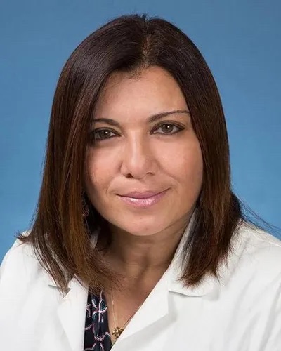
    Maie A. St. John

    Maie A. St. John, MD, PhD, FACS

    Andelot Professor of Laryngology and Otology · Professor of Oncology
    Director, Department of Otolaryngology–Head and Neck Surgery
    Johns Hopkins University School of Medicine

    Dr. St. John is a head and neck surgical oncologist whose research centers on the mechanisms of tumor progression and metastasis in head and neck squamous cell carcinoma — and on translating those mechanisms into tools a surgeon can pick up. Her NIH- and NCI-funded work spans intraoperative optical imaging, targeted local delivery of therapeutics, the tumor microenvironment, and patient-derived models.

    She was named Director of the Johns Hopkins Department of Otolaryngology–Head and Neck Surgery in 2025, after serving as Professor and Chair of the Department of Head and Neck Surgery at UCLA.

    • 2025–Andelot Professor & Director, Otolaryngology–Head and Neck Surgery, Johns Hopkins
    • 2017–25Professor & Chair, Head and Neck Surgery, UCLA
    • 1999MD, Yale School of Medicine (Medical Scientist Training Program)
    • 1998PhD, Yale School of Medicine
    • 1992BS, Stanford University
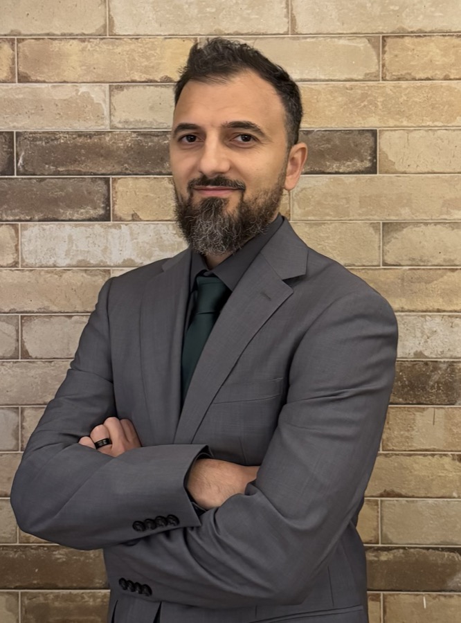
    Yazeed Alhiyari

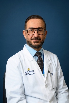
    Yazeed Alhiyari, PhD

    Senior Scientist
    Department of Otolaryngology–Head and Neck Surgery
    Johns Hopkins University School of Medicine

    A biomedical physicist working where optics, molecular biology, and computation meet. Dr. Alhiyari has been central to the development of Dynamic Optical Contrast Imaging — building and validating the platform across oral cavity margins, parathyroid localization, thyroid pathology, perineural invasion, and vocal fold scar, and more recently pairing it with machine-learning segmentation.

    His work extends past imaging: he led the design of a modular polymer platform for local delivery of head and neck cancer therapy, and contributed to the lab's work on sensory nerve signaling in the tumor microenvironment and on tissue-engineered vocal fold replacement. His earlier research, in radiation oncology, examined metabolism and Notch signaling in cancer stem cells.

    • PresentSenior Scientist, Otolaryngology–Head and Neck Surgery, Johns Hopkins
    • PriorAssistant Project Scientist, Head and Neck Surgery, UCLA
    • 2014PhD, Biomedical Physics, University of California, Los Angeles
    • EarlierMS, Molecular Pathology · bioinformatics and molecular genetics research

    Lab members

    Postdocs · residents · graduate students · research staff

    Zeena Mestari

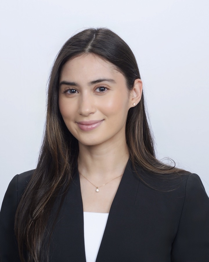
    Zeena Mestari

    Medical Student, David Geffen School of Medicine at UCLA · Research Associate, Johns Hopkins University

    Zeena's work has focused on biologic therapies and clinical trials. With a background in applied behavior analysis and medical device development, her research interests include emerging therapies and diagnostic technologies aimed at improving patient outcomes and quality of life.

    Dina Garmroudi

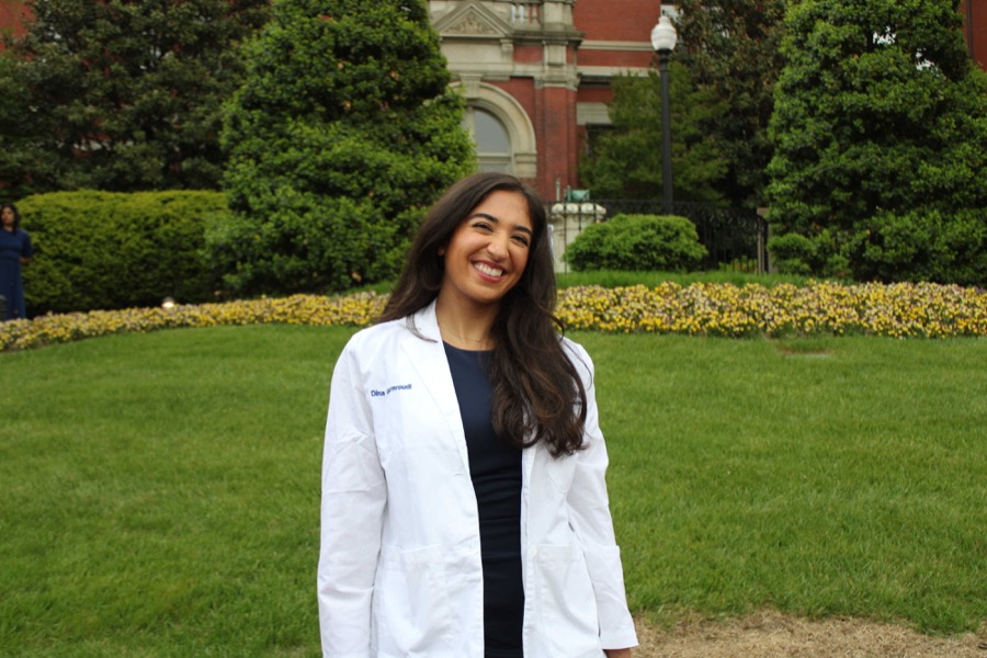
    Dina Garmroudi

    Medical Student, Johns Hopkins University School of Medicine

    Originally from Maryland, Dina completed her undergraduate and M.P.H. degrees at Yale University and is interested in pursuing head and neck cancer surgery. She hopes to research and help develop preventative and screening efforts for head and neck cancer, and to improve patient outcomes. Outside the lab she enjoys running, biking, exploring cafés, and traveling.

    +

    Postdoctoral fellow

    Add member here
    +

    Graduate student

    Add member here
    Alumni

    Add former lab members here, with where they went next.

    Join

    We look for people who want the hard problem

    The lab is deliberately mixed — surgeons, engineers, biologists, and students who have not decided yet. If you want your work to end up in an operating room, write to us.

    Postdoctoral fellows

    Optical engineering, cancer biology, or drug delivery. Send a CV, a short statement, and two references.

    Graduate students

    Rotations welcome through affiliated Hopkins programs. Tell us which program pulls at you, and why.

    Residents & clinical fellows

    Protected research time within Otolaryngology–Head and Neck Surgery. Projects scale to the time you have.

    Undergraduates

    A small number of positions each term, for students ready to commit real hours rather than a summer cameo.

    Contact

    The margin you cannot see is the one that comes back.

    • Email
      lab@stjohnlab.org
    • Department
      Department of Otolaryngology–Head and Neck Surgery
      Johns Hopkins University School of Medicine
    • Address
      Ross Research Building, Room 715
      720 Rutland Avenue
      Baltimore, MD 21205
    • Elsewhere